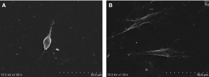
Figure 4.

(A) Scanning electron microscopy showed that cells cultured in the control medium had a long and thin spindle shape, with tiny, short and less bumps stretching out of the cellular surface. (B) By contrast, cells cultured in the PRF medium were larger and showed a fusiform shape, with radial, longer and many more bumps stretching out of the cellular surface.
