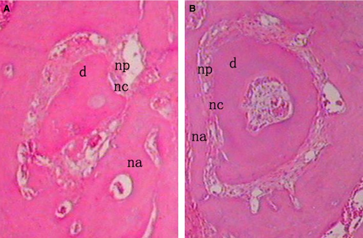
Figure 8.

In the PRF group (A), little new cementum could be observed at the margin of the defect and the PRF + cells group (B) showed a thin layer of new cellular cementum covering the root‐denuded surface at 24 days after surgery. All of the new cementum was cellular cementum, and cracks between the new cementum and the root dentin were observed in some cases. The regenerated PDL fibres separating the new bone from the new cementum was disordered and not perpendicular to the root surface in the PRF + cells group (B) and those in the PRF group (A) were sparse and loose without periodontal fibre bundle formation at 24 days after surgery (×200). d, dentin; np, new periodontal ligament; na, new alveolar bone; nc, new cementum.
