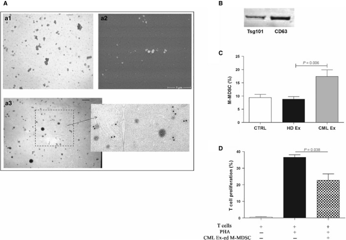
Figure 4.

CML exosomes promote the generation of M‐MDSC. (A) a1: Representative TEM image of CML serum exosomes (Ex). The exosomes show a characteristic ‘deflated football‐shaped’ of 60–100 nm in size (Bar = 120 nm). a2: A S.E.M. image of CML exosomes at high magnification (× 30,000). a3: The exosomes are positive for exosomal marker CD81 (Bar = 120 nm). Right panel: boxed area shown at higher magnification. (B) Western blot analysis of protein extracted from exosomes. (C) An increase in the percentage of CD14+/HLA‐DR − cells was observed in vitro after incubation of HD monocytes with CML exosomes (P < 0.05). Results represent the means of four independent experiment; error bars denote S.D. (D) Suppressive activity of CML exosomes‐educated M‐MDSC (CML Ex‐ed M‐MDSC) was evaluated in coculture experiments with CFSE‐labelled autologous T lymphocytes. Mean frequency of CD3+ CFSE dim cells ± S.D. from four independent experiments in duplicate is shown.
