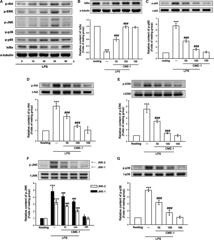
Figure 2.

Time‐course analysis of lipopolysaccharide (LPS)‐activated signalling molecules and effects of CME‐1 on LPS‐induced IκBα degradation and the phosphorylation of p65, Akt, ERK, JNK and p38 in RAW 264.7 cells. (A) RAW 264.7 cells were treated with LPS (1 μg/ml) for the indicated time. IκBα degradation and p65, Akt, ERK, JNK and p38 phosphorylation were determined using an immunoblotting assay as described in the Methods. (B–G) RAW 264.7 cells were treated with PBS (resting group) or CME‐1 (25–100 μg/ml) for 20 min, followed by LPS (1 μg/ml) for 30 min. IκBα degradation and the phosphorylation of p65, Akt, ERK, JNK and p38 were evaluated using immunoblotting. Data are presented as the mean ± S.E.M. (n = 3). ***P < 0.001, compared with the resting group; ### P < 0.001, compared with the LPS group.
