
Figure 5.
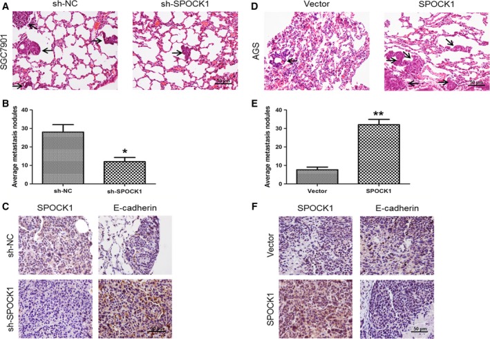
Influences of SPOCK1 silencing and overexpression on the metastasis of gastric cancer cells in vivo. (A,D) Lung metastatic nodules (indicated by black arrows) were histologically observed through HE‐stained samples. (B,E) The number of average lung metastatic nodules was calculated. (C,F) Expressions of SPOCK1 and E‐cadherin in lung metastatic nodules were determined by immunohistochemistry. *P < 0.05, **P < 0.001. Scale bar = 50 μm.