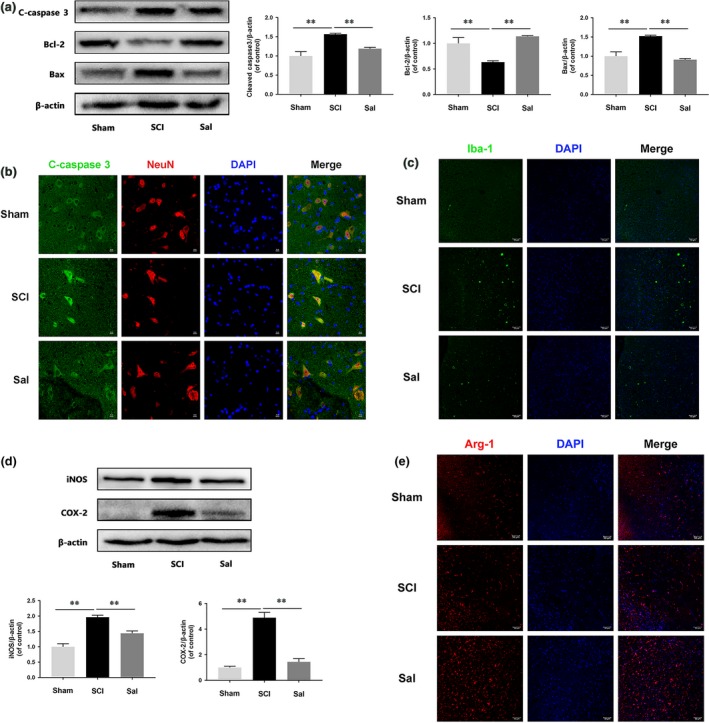
Figure 2.

Sal prevents neuronal apoptosis and regulates microglia/macrophage polarization (A) Western blot analysis of cleaved caspase 3, Bcl‐2 and Bax expression in each group of rats. Sal evidently prevented SCI‐induced neuronal apoptosis. (B) Double‐staining for cleaved caspase 3 (green)/NeuN (red) in sections of injured spinal cord tissue from each group of rats (scale bar: 10 μm). (C) Immunofluorescence staining for Iba‐1 in injured spinal cord tissue from each group rats (scale bar: 50 μm). Sal significantly reduced the numbers of M1 microglia after SCI. (D) Representative Western blots of and quantitative data for iNOS, COX‐2 and β‐actin expression in each group of rats. Inflammatory mediator release was suppressed by Sal treatment. (E) Immunofluorescence staining for Arg‐1 in sections of injured spinal cord tissue from each group rats (scale bar: 50 μm). Sal significantly increased M2 cell numbers after SCI. Densitometric analysis of all Western blot bands, whose densities were normalized to those of β‐actin. Data are presented as the mean ± S.D., n = 3 independent experiments. Significant differences between groups are indicated as *P < 0.05 and **P < 0.01.
