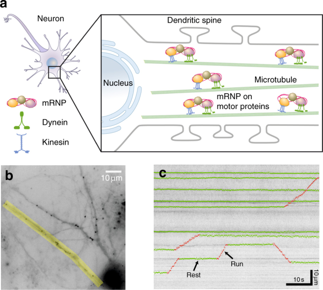
Fig. 1.

Schematic and experimental data showing motor-driven transport of mRNPs in a neuron. a An ensemble of mRNP complexes are transported by kinesin and dynein motor proteins along microtubules in the dendrite. In each dendrite, there are multiple microtubule tracks and thus several mRNP particles are efficiently transported at the same time to the target sites where they are localized. b A live-cell image showing fluorescently labeled β-actin mRNP complexes in a hippocampal neuron. Time-lapse images were taken with a time interval of t0 = 0.1 s for the overall observation time of T = 60 s. The region of interest for analysis is marked in yellow. Scale bar, 10 μm. c A kymograph for an ensemble of mRNP particles obtained from an image similar to the yellow area in (b). Horizontal and vertical axes correspond to the elapsed time and the distance along the dendrite, respectively. For a few trajectories, their two distinct dynamic modes, rest and run, are denoted in green and red, respectively. The kymographs exhibit constant small-length scale fluctuations (see Supplementary Fig. 1 for more information). Scale bars, 10 s and 10 μm, respectively
