Figure 7.
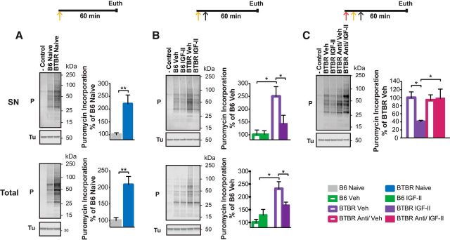
IGF-II reduces overactive translation in the hippocampus of BTBR mice. Experimental timelines are shown above graphs. Active translation was measured by puromycin incorporation, which was injected bilaterally into the dorsal hippocampus (yellow ↑). Immediately after the puromycin injection mice received a subcutaneous injection (↑) of IGF-II or vehicle (Veh; B) in the presence or absence of anti-IGF-II receptor blocking antibody (C). Representative blots are shown beside their respective grouped data. Each relative value was normalized against β-tubulin (Tu) detected on the same blot. All data are expressed as the mean (±SEM) of B6 naive (A), B6 vehicle-injected (B), or BTBR vehicle-injected (C) mice. n = 6–8 per group. −Control represents the labeling of control samples without puromycin. A, B, Two-way ANOVA followed by Tukey post hoc tests. C, One-way ANOVA followed by Tukey post hoc tests. *p < 0.05, **p < 0.01. A, Western blot analyses of puromycin (P) incorporation in SN (top) and total protein (bottom) extracts obtained from dorsal hippocampi of untrained B6 and BTBR mice killed 60 min after P injection. B, Western blot analyses of P incorporation in SN (top) and total protein (bottom) extracts obtained from dorsal hippocampus of B6 and BTBR mice injected subcutaneously with either Veh or IGF-II and killed 60 min after the injection. C, Western blot analyses of P incorporation in SN protein extracts obtained from dorsal hippocampi of BTBR mice injected bilaterally into the dorsal hippocampus with an IGF-IIR functionally blocking antibody (Anti; red ↑) or IgG immediately before a subcutaneous injection of Veh or IGF-II and killed 60 min after the subcutaneous injection. For detailed statistical analyses see the Extended Data table (Fig. 7-1).

