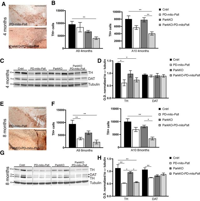
Figure 2.
Degeneration of dopaminergic neurons in substantia nigra and loss of striatal axonal projections. A, E, Representative images of the midbrain sections immunostained to identify TH+ neurons for PD-mito-PstI and ParkinKO-PD-mito-PstI mice at 4 (A) and 8 (E) months of age. Scale bar, 500 μm. B, F, Graph representing the stereological quantification A9 and A10 TH+ cells (n = 4–5/group) in 4-month-old (B) and 8-month-old (F) mice. C, D, Western blots showing levels of TH and DAT in 4-month-old mice striata (C) and relative quantification (D). G, H, Western blots showing levels of TH and DAT in 8-month-old mice striata (G) and relative quantification (H). Molecular weights are indicated on the left of the gels. Error bars ± SEM. n = 5/group. *p < 0.05, **p < 0.01.

