Figure 3.
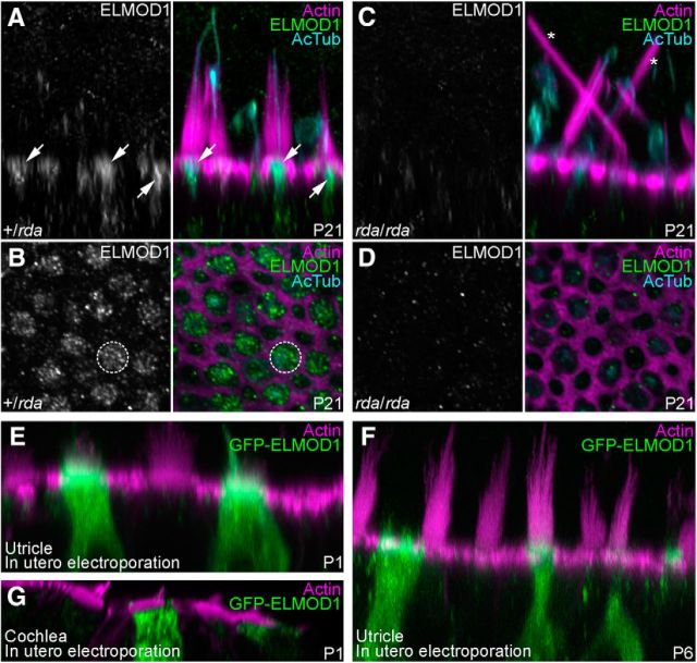
Localization of ELMOD1 in utricle hair cells. A–D, Confocal images of utricle hair cells from P21 +/rda (A, B) or rda/rda (C, D) mice. Images were x-z reslices (A, C) to show ELMOD1 distribution at the apical region of hair cells (arrows) or x-y confocal sections (B, D) to show ELMOD1 at the cuticular plate level (dashed circle). No ELMOD1 signal is seen in rda/rda utricles (C, D); asterisks indicate giant fused stereocilia. Utricles were labeled with phalloidin (magenta) and stained with anti-acetylated tubulin (cyan) and anti-ELMOD1 (green) antibodies. E–G, Confocal images of vestibular (E, F) or cochlear (G) hair bundles from mice electroporated in utero with a GFP-ELMOD1 construct at E11.5 and dissected at P1 (E, G) or P6 (F). Utricles and cochleae were labeled with phalloidin (magenta) and stained with anti-GFP antibody (green) to amplify GFP-ELMOD1 fluorescence. Panel full widths: A–D, 25 μm; E, 40 μm; F, G, 50 μm.

