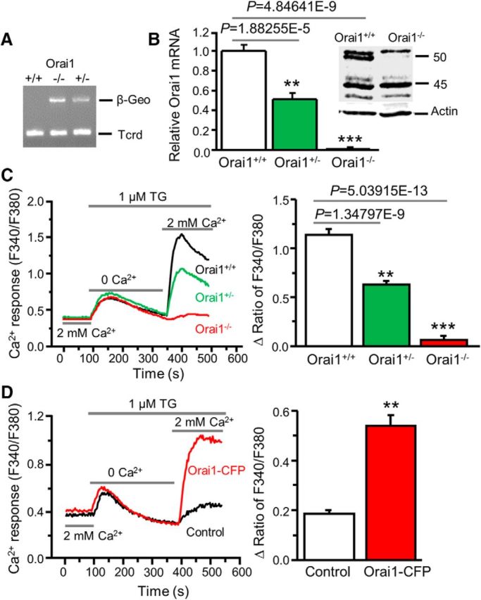
Figure 4.

Orai1 deficiency abolishes SOCE in dorsal horn neurons. A, β-Geo expression in Orai1 knock-out (Orai1−/−), heterozygous (Orai1+/−), and wild-type (Orai1+/+) mice. T-cell receptor delta chain (Tcrd) is a control for presence of DNA. B, Orai1 mRNA expression (relative to GAPDH and normalized to Orai1+/+) in Orai1+/+, Orai1+/−, and Orai1−/− mice (n = 5–6 mice, F = 94.5, p values indicated in graph). Inset, Western blot of Orai1 protein extracted from spinal cord tissues of Orai1+/+ and Orai1−/− mice. C, TG-induced Ca2+ entry in Orai1+/+, Orai1+/−, and Orai1−/− dorsal horn neurons (n = 26–34 neurons, F = 44.1, p values indicated in graph). D, TG-induced Ca2+ entry in Orai1−/− neurons transfected with Orai1+/+ Orai1-CFP or GFP (as control; n = 17–19 neurons, df = 16, t = 7.58176, p = 1.10506E-6). Values represent mean ± SEM; **p < 0.01, ***p < 0.001 compared with Orai1+/+ or control neurons by one-way ANOVA or the paired t test.
