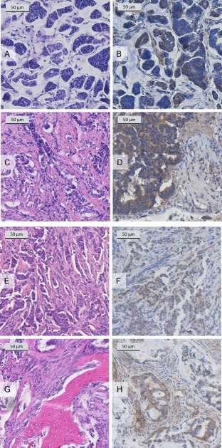
Figure 5.

Immunostaining of specimens from breast cancer patients. Mucinous, grade 2 breast cancer of a 45‐year‐old female patient (ER positive, PR weakly positive, Her2 negative; (A) HE stain of the primary carcinoma (×10 objective magnification); (B) hypo‐BSP stain of the primary carcinoma with rat monoclonal antibody IDK‐1 (×20 objective magnification) showing moderate local expression; (C) HE stain of metachronous (7 years) spinal metastasis (thoracic vertebrae 3 + 4, ×4 objective magnification); (D) hypo‐BSP stain of a metachronous (7 years) spinal metastasis with rat monoclonal antibody IDK1 (×40 magnification) showing diffuse distribution of moderate cytoplasmic hypo‐BSP expression. Invasive, grade 2 ductal breast cancer of a 47‐year‐old female patient (ER strongly positive, PR strongly positive, Her2 negative; (E) HE stain of the primary carcinoma (×4 objective magnification); (F) hypoglycosylated BSP stain of the primary carcinoma with rat monoclonal antibody IDK1 (×4 objective magnification) showing focal, moderate cytoplasmic expression; (G) HE stain of a synchronous spinal metastasis (thoracic vertebra 6, ×20 objective magnification); (H) hypoglycosylated BSP stain of a synchronous spinal metastasis with rat monoclonal antibody IDK1 (×20 objective magnification) showing diffuse distribution of moderate to strong cytoplasmic expression.
