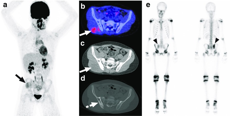
Fig. 2.
Non-FDG-avid sclerotic bone marrow involvement of the right posterior iliac crest in a 10-year-old boy with Ewing sarcoma. Coronal maximum intensity projection FDG-PET (a), axial FDG-PET/CT (b), and low-dose CT with soft tissue window settings (c) show the slightly FDG-avid primary tumor in the right gluteal muscles (continuous arrow). Although no increased FDG uptake is seen in the right posterior iliac crest (b), low-dose CT with bone window settings shows extensive sclerosis in the right posterior iliac crest (dashed arrow). Blind BMB of the right posterior iliac crest showed involvement with Ewing sarcoma. Of interest, bone scintigraphy (e) that was performed before FDG-PET/CT also showed pathological activity in the right posterior iliac crest (arrowheads)

