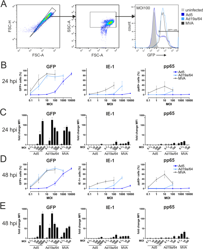
Figure 3.
MVA and Ad19a/64 efficiently transduce dendritic cells. MoDCs from 3 different HCMV seronegative blood donors were infected at varying MOIs with the indicated vectors and intracellular presence of the transgenes GFP, IE-1 and pp65 was quantified via flow cytometry after 24 (B,C) and 48 hours (D,E). Panel A shows the gating strategy as well as a representative histogram from one donor 48 hours after transduction of moDCs with GFP expressing vectors at MOI 100. Summarized results are given as the median percentage of cells positive for each antigen (B,D) with error bars representing the standard deviation. Median fluorescence intensity (MFI) values were normalized to the signals obtained from uninfected cells with bars representing the mean and standard deviation of values from all donors (C,E). Histograms displaying IE-1 and pp65 expression individually for each donor are shown in Supplementary Figure 2.

