Figure 6.
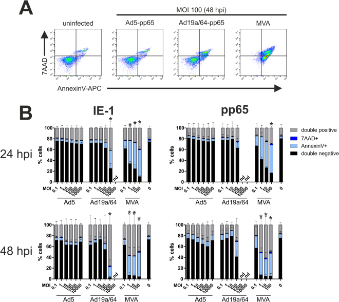
MVA causes the highest amount of cell death in dendritic cells. MoDCs from 3 different HCMV seronegative blood donors were transduced at varying MOIs with the indicated vectors. 24 or 48 hpi, cells were stained with AnnexinV-APC and 7AAD before flow cytometric analysis. Panel A shows representative plots 48 hours after transduction with the indicated vectors at MOI 100. The results from all donors are summarized in (B). Bars show the mean and standard deviation (SD) of values from all donors (nd: not determined). Asterisks indicate conditions where the fraction of viable (double negative) cells was significantly (p < 0.05, Bonferroni-corrected t-test) reduced as compared to the uninfected control.

