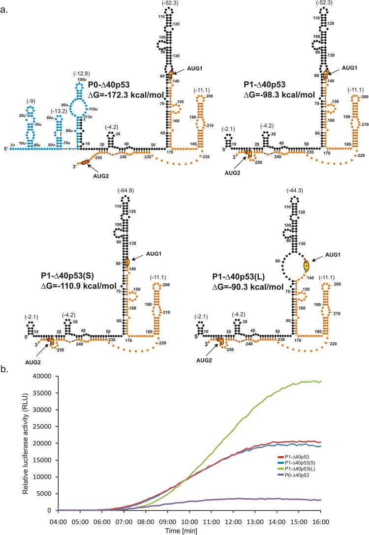
Figure 4.
Secondary structure models of 5′-terminal regions of mRNA constructs with two initiation codons AUG1 and AUG2 and kinetics of their in vitro translation followed by a luciferase reporter gene assay. (a) Secondary structure arrangement of the 5′UTRs of constructs: P0-Δ40p53, P1-Δ40p53, P1-Δ40p53(S) and P1-Δ40p53(L). The predicted ΔG values for each 5′UTR and for selected structural motifs are shown. Colours denote parts of the 5′-terminal region of p53 mRNA as described in the legend to Fig. 2. (b) The relative luciferase activity (RLU) was measured during translation of each mRNA construct using a luminometer in 1 s periods with 9-second intervals, for 16 minutes.

