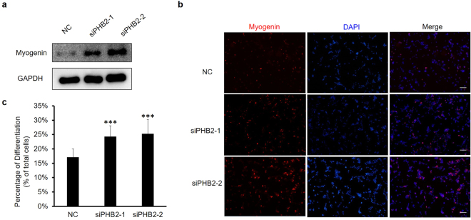
Figure 3.
PHB2 knockdown promotes RD cell differentiation. (a) Detection of myogenin by Western blot. RD cells were transfected with siRNA and the growth medium was replaced 6 hours later. The next day, the medium was replaced with the differentiation medium (2% calf serum, H-DMEM). Cell extracts were subjected to immunoblotting with indicated antibodies, and GAPDH was detected as a loading control. Full-length blots are presented in Supplementary Fig. S1. (b) Immunofluorescence staining of myogenin in RD cells. Forty-eight hours after transfection, RD cells were fixed and subjected to immunostaining for myogenin (red). The nuclei of cells were counterstained with DAPI (blue). Scale bar 200 μm. Results are representative of three independent experiments. (c) Quantitative analysis of myogenin in panel B. Data are presented as the mean ± S.D. (n = 3), *P < 0.05, **P < 0.01, ***P < 0.001, two-tailed Student’s t test.

