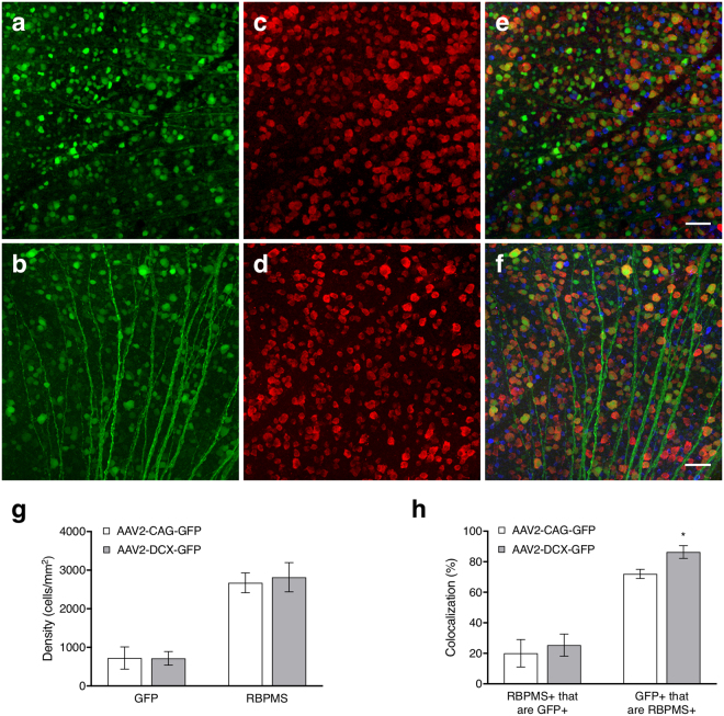
Figure 3.
Cellular labelling in flat-mount retinas with the two AAV2 vectors. Panels (a,c and e) are micrographs from an animal that received an injection of the AAV2-CAG-GFP vector while panels (b,d and f) are from an animal that received an injection of the AAV2-DCX-GFP vector. (a,b) Show GFP labelling via intravitreal injection of AAV2-GFP vector (green); (c,d) immunohistochemical labelling of RGCs with RNA binding protein with multiple splicing (RBPMS, red) and (e,f) respective merged images with choline acetyltranferase (ChAT, blue). Scale bars = 50 μm. (g) Density of GFP positive cells from an AAV intravitreal injection and RBPMS positive cells from immunohistochemical labelling. (h) RBPMS+ cells that are GFP+ indicates the proportion of RGCs labelled by GFP and GFP+ cells that are RBPMS+ indicates the specificity of the AAV vector to RGCs. Histograms show means and error bars show 95% confidence intervals; *p < 0.05; n = 6.

