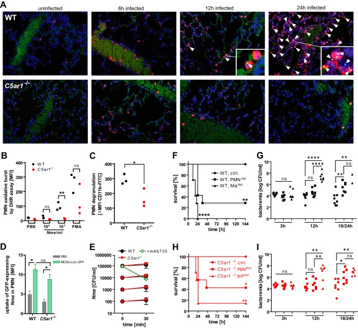
FIG 4 .
Role of phagocytes in N. meningitidis sepsis in WT and C5ar1−/− mice. (A) Immunofluorescence microscopy (×60 magnification) of tissue sections of lung at indicated time points after intraperitoneal infection with 105 CFU N. meningitidis MC58. Blue, nuclei; red, neutrophil elastase; green, N. meningitidis (arrowheads). Background data in the green channel stem from erythrocytes, indicating the position of blood vessels. Insets are enlargements of points of interest from the same image. (B) Oxidative burst of Ly6Ghi neutrophils (n = 3) assayed by DHR123 assay in lepirudin-treated whole blood infected with 105 or 107 CFU per ml of N. meningitidis MC58. The positive control was PMA (100 nM). ns, not significant; **, P < 0.01 (in unpaired, two-tailed Student’s t test). (C) Neutrophil degranulation measured as the difference in levels of CD11b surface expression between infected (107 CFU/ml) and uninfected lepirudin-treated whole-blood samples from WT and C5ar1−/− mice after 1 h of incubation. Neutrophils were gated as Ly6Ghi cells and CD11b stained with clone M1/70. *, P < 0.05 (in unpaired, two-tailed Student’s t test). (D) Uptake of N. meningitidis MC58Δcsb-GFP by Ly6Ghi neutrophils in ex vivo infection of lepirudin-treated whole blood (means ± SEM of the geometric mean of GFP fluorescence; n = 3). ns, not significant, *, P < 0.05 (in one-way ANOVA with Bonferroni’s post hoc test). (E) Ex vivo N. meningitidis survival at different inocula in lepirudin-treated whole blood of WT and C5ar1−/− mice (means of CFU per milliliter ± SEM; n = 3). As a positive control for N. meningitidis killing, 1 µg/ml of anti-serogroup B mouse monoclonal antibody mAb735 was added. (F and H) Survival of n = 7 to 8 WT and C5ar1−/− mice, respectively, infected with 104 CFU of N. meningitidis MC58 after depletion of monocytes/macrophages (clodronate liposomes) or neutrophils (RB6-8C5) or the control (PBS). The experiment was conducted in a blind manner for depletion treatment. *, P < 0.05; **, P < 0.01; ***, P < 0.001; ****, P < 0.0001 (in Mantel-Cox analysis relative to control). (G and I) N. meningitidis counts in blood of mice in panels G and I. ns, not significant; *, P < 0.05; **, P < 0.01; ****, P < 0.0001 (in one-way ANOVA, applying Bonferrroni’s post hoc test).

