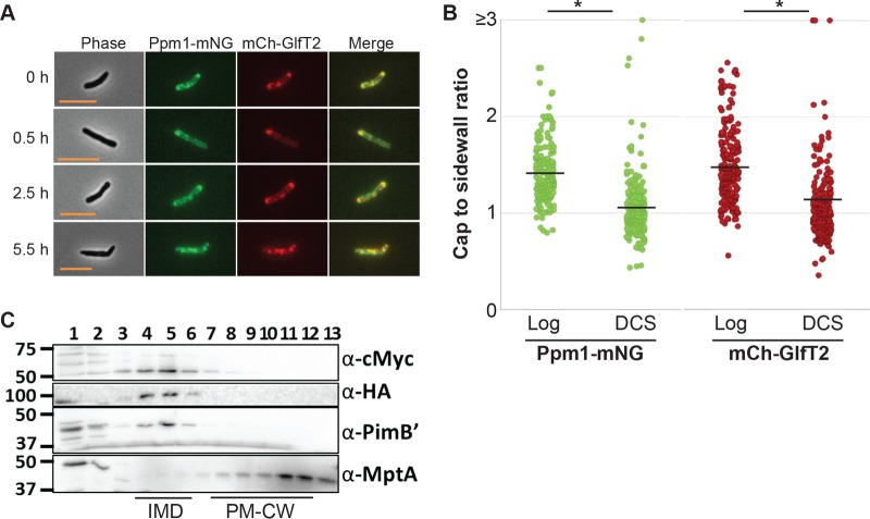
FIG 3 .
Inhibition of PG synthesis by DCS leads to reorganization of the IMD. (A) Fluorescence microscopy images of the dual IMD marker strain expressing HA-mCherry-GlfT2 (mCherry-GlfT2) and Ppm1-mNeonGreen-cMyc (Ppm1-mNG). DCS treatment led to the relocalization of the IMD from polar to sidewall enrichment. Scale bar, 5 µm. (B) The cap-to-sidewall ratio of two marker proteins in cells treated with DCS for 6 h compared with cells before treatment (log), quantitatively demonstrating reorganization from the polar cap to sidewall enrichment. The black lines indicate the averages of 218 cells. (C) Western blotting detection of IMD proteins (Ppm1-mNeonGreen-cMyc, 59 kDa; HA-mCherry-GlfT2, 100 kDa; PimB′, 42 kDa) and the PM-CW protein (MptA, 54 kDa) which were separated by sucrose density gradient sedimentation, illustrating enrichment in IMD fractions after 8 h of DCS treatment. *, P < 0.001.

