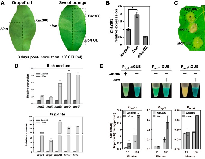
FIG 2 .
Lon negatively regulates bacterial virulence. (A) Grapefruit and sweet orange leaves were inoculated with wild-type strain 306 (Xac306), the Δlon mutant, and a Lon-overexpressing strain (Δlon OE) with a needleless syringe. The bacterial titer of the inoculum was adjusted to 106 CFU ml−1. The canker symptoms induced by the corresponding strains were photographed at 3 days postinoculation. The experiments were repeated three times with comparable results, and only one representative leaf is shown. (B) CsLOB1 expression levels in grapefruit leaves inoculated with wild-type strain 306, the Δlon mutant, and the Δlon OE strain. CsLOB1 expression was monitored by qRT-PCR at 48 h postinoculation. The mean values ± the standard deviations (n = 3) are plotted. The asterisk indicates a statistically significant difference (P < 0.01, Student t test). (C) Fully expanded 4-week-old N. benthamiana leaves were inoculated with wild-type strain 306, the Δlon mutant, and the Δlon OE strain at a concentration of 108 CFU ml−1. HRs were observed and photographed at 1 week postinoculation. The experiments were repeated three times with comparable results, and only one representative leaf is shown. (D) Comparison of relative hrp/hrc gene expression in wild-type strain 306 and the Δlon mutant in rich medium (top) and in planta (bottom). RNA samples were extracted from bacterial cells either grown in NB medium to stationary phase or recovered from plant leaves at 4 days postinoculation. Relative hrpG, hrpX, hrcQ, hrcU, and hrpB1 expression was monitored by qRT-PCR. The mean values ± the standard deviations (n = 3) are plotted. (E) The hrpB1, hrcU, and hrcQ promoters were fused with a GUS reporter gene, and the derivative plasmids were transferred into the wild type and the Δlon mutant. Transformants were grown in rich medium overnight and stained with X-Gluc for 10 min. The experiments were repeated three times with comparable results, and only one tube is presented. Quantification of GUS activity was performed with p-nitrophenyl-β-d-glucuronide as the substrate. The mean values ± the standard deviations (n = 3) are plotted.

