Figure 3.
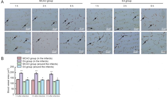
Effect of EA on quantity of blood-flowing vessels in and around cerebral infarcts in MCAO rats (lectin staining).
(A) Blood vessel staining around (upper) and within (lower) infarcts (original magnification, 100×). Arrows indicate blood vessels. Scale bars: 20 μm. (B) Blood vessel quantity in and around infarct lesions. Data represent mean ± SEM (n = 10). Independent samples t-test was used if data met the normal distribution and homogeneity of variance. Separate variances t-test was used if data met the normal distribution but unequal variance. Wilcoxon rank test for independent samples was used for skewed distribution data. *P < 0.05, **P < 0.01, vs. MCAO group. MCAO: Middle cerebral artery occlusion; EA: electroacupuncture; h: hour(s).

