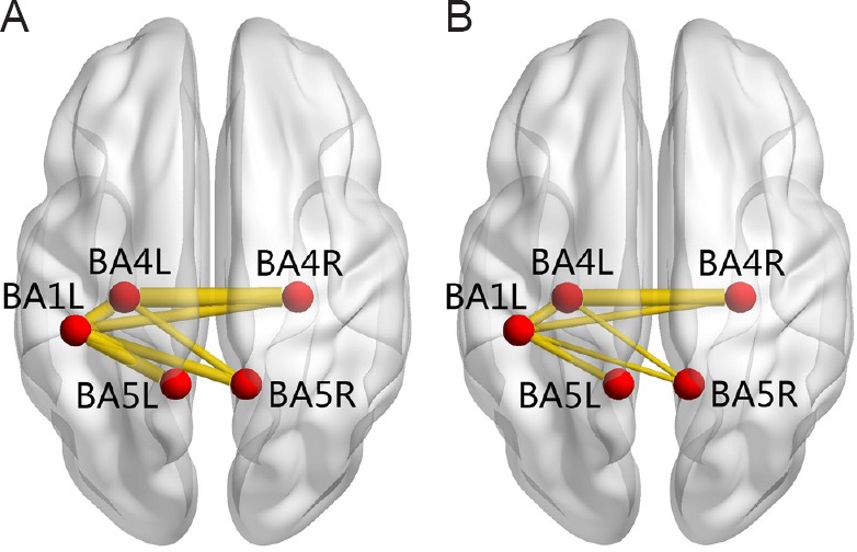
Figure 1.

Anatomic replicas show the decreased functional connectivity of sensorimotor brain areas in patients with SCI compared with healthy subjects.
(A) Normal functional connectivity in healthy subjects; (B) decreased functional connectivity in patients with spinal cord injury. The red nodes represent the seed areas and significantly changed areas of functional connectivity. The yellow lines represent decreased functional connectivity in spinal cord injury patients relative to healthy subjects. BA1L: Left primary somatosensory cortex; BA4L: left primary motor cortex; BA4R: right primary motor cortex; BA5L: left somatosensory association cortex; BA5R: right somatosensory association cortex.
