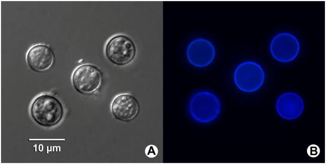
Fig. 2.
Photomicrographs of Cyclospora macacae oocysts isolated from feces of rhesus monkeys (Macaca mulatta) in China. (a) Oocysts under differential interference contrast microscopy of wet mount. (b) Typical blue autofluorescence of the oocysts is observed under epifluorescence microscopy using a 330–380-nm ultraviolet excitation filter

