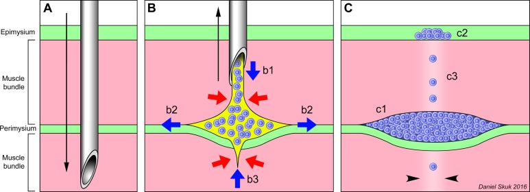
Figure 9.
Interpretation of the early location of the injected cells. (A) Penetration (black arrow) of the injection needle through the muscle bundles (pink) and connective tissue (green). (B) Delivery of the cell suspension (blue = grafted cells, yellow = saline) during the needle withdrawal. Red arrows represent the compression exerted by the tissue on the cell suspension, and blue arrows represent the cell suspension displacements into the muscle. Even if the cell suspension is homogeneously delivered (b1) during the removal (black arrow) of the needle, it does not remain in the needle’s trajectory but rather split the boundary between the perimysium and the muscle bundle (b2). The cell suspension may be expelled from the muscle bundles traversed by the needle (b3). (C) The final result, after saline absorption, is that most grafted cells are compacted in the periphery of muscle bundles (c1), some in the epimysium (c2), and only a few remain in the needle trajectory inside the fascicles (c3; the damage of the needle is represented by paler color, between arrowheads).

