Fig. 2.

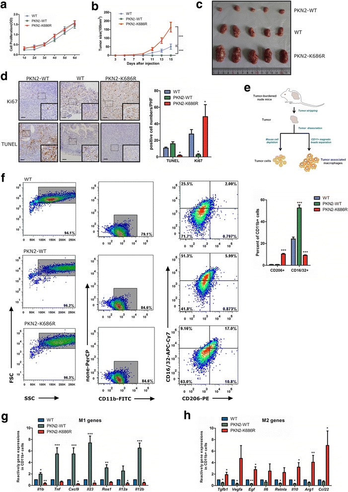
PKN2expression inhibits tumorformationand decreased macrophages polarizing towards the M2 type in the xenograft tumor. a HCT116 cells were stably infected with vector (WT), PKN2-WT, PKN2-K686R lentivirus and cultured for 1-6 days. Cell proliferation was detected by CCK8 assay. b Tumorigenesis assay of Balb/c nude mice subcutaneously injected with PKN2-WT/PKN2-K686R transduced or wild-type HCT116cells (n = 10). ***, P < 0.001 versus WT. c Representative photos of tumors from mice of the various groups. d TUNEL assay &IHC staining of Ki67 in tumor tissues in mice xenograft model and positive cell numbers per high field were counted.*, P < 0.05 versus WT. e Schematic picture on the procedure for separation of Tumor cells &TAM. f CD11b+ macrophages were separated from murine tumor tissues using CD11b magnetic beads. Surface expression of CD16/32 and CD206 was detected in CD11b+ macrophages usingflow cytometry.The percent of CD16/32+ or CD206+ cells in CD11b+macrophages were assayed.***, P < 0.001 versus WT. g Relative gene expression of M1 marker (Il1b, Tnf,Cxcl9, Il23, Ros1,Il12a, and Il12b) and M2 marker(Tgfb1, Vegfa, Egf, Il6, Retnla, Il10, Arg1 and Ccl22) (h) in the tumor tissues of mice.*, P < 0.05; **, P < 0.01; ***, P < 0.001 versus WT
