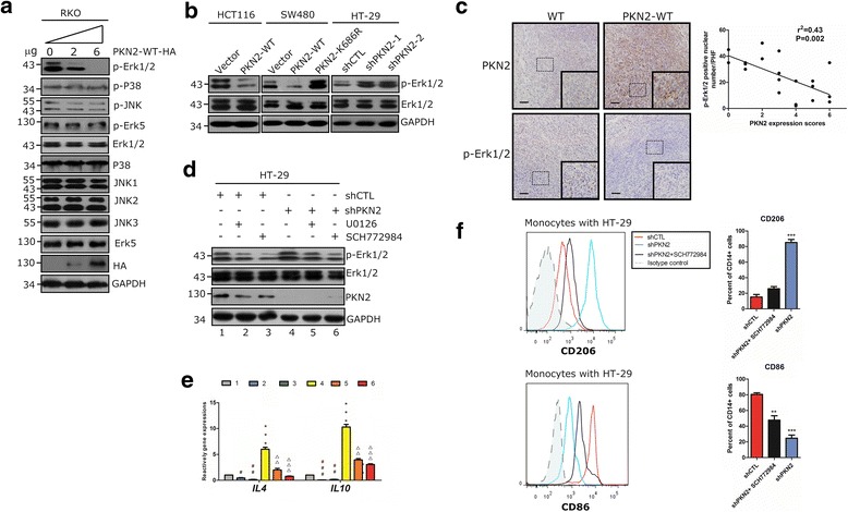
Fig. 5.

PKN2 negatively regulates Erk1/2. a RKO cells were transfected with 0, 3 or 6 μg PKN2-WT-HA.Western blotting was used to detect the indicated proteins. b Stable clones of SW480, HCT116 and HT-29 cells (as indicated in Fig. 3) were detected for the expression of p-Erk1/2, Erk1/2 and GAPDH using western blotting. c IHC staining of PKN2 and p-ERK1/2 in the tumor tissues of mice xenograftmodels. The correlation between p-Erk1/2 positive number per high field and the PKN2 expression score was explored. d HT-29cells were stably transfected with shCTL or shPKN2 and treated with solvent, SCH772984 (1 μM) or U0126 (1 μM) for 1 h. Western blotting was used to detect the indicated proteins. e HT-29 cells were treated as indicated in (d). IL4 and IL10 gene expression was detected by RT-PCR.***, P < 0.001 versus lane 1. #, P < 0.05; ##, P < 0.01; ###, P < 0.001 versus lane 1. △△, P < 0.01; △△△, P < 0.001 versus lane4. f Human CD14+ monocytes were cocultured with stably transfected shCTL or shPKN2 HT-29 cells treated with SCH772984 (50 nM) or solvent for 4 days. Surface expression of CD206 and CD86 in differentiated macrophages was detected using flow cytometry.**, P < 0.01; ***, P < 0.001 versus shCTL
