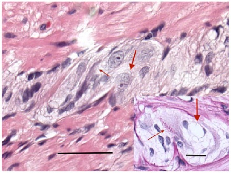
Figure 2.

Myenteric ganglion with neurons showing vacuolated cytoplasm (thick arrow) and a shrunken, darker, amphophilic, pre-apoptotic neuron also with a few vacuoles (thin arrow) (hematoxylin and eosin stain) (bar: 50 µm). Insert: large accumulation of lipofuscin granules within a neuron (thin arrow). Thick arrow points to a lacuna after lysis of a necrotized neuron with the nuclei of two glial cells (periodic acid-Schiff-diastase stain) (bar: 20 µm).
