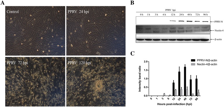
Figure 2.

PPRV replication and nectin-4 expression in EECs. A Morphological changes in infected EECs at the indicated time points (magnification, ×100). B PPRV-infected cells were collected for western blotting with anti-PPRV-N and anti-nectin-4 antibodies at the indicated time points. β-Actin was detected as the loading control. Representative results are shown and similar results were obtained in three independent experiments. C Analysis of the relative levels of nectin-4 and PPRV-N in infected EECs. The optical densities for the nectin-4, PPRV-N, and β-actin protein bands were measured with densitometric scanning, and the ratios of nectin-4/β-actin and PPRV-N/β-actin were calculated. The data are expressed as the mean ± SD of three independent experiments.
