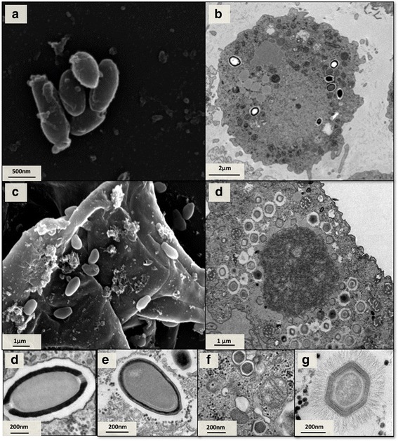
Fig. 4.

Electron microscopy images of viruses isolated. SEM of Cedratvirus isolated from sewage farm of MG (a) TEM (b) and SEM (c) of Pandoravirus isolate from Mergulhão sewage creek. TEM of Mimivirus isolated from Antarctica (c) TEM of Cedratvirus isolated from sewage farm of MG (d) TEM of Pandoravirus isolate from Bom Jesus sewage creek (e) marseillevirus isolated from Bom Jesus sewage creek (f). TEM mimivirus particle detail that was isolated from Antarctica (g). Scale Bars: (a-d) 500 nm; (e) 50 nm
