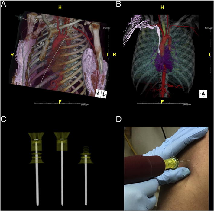
Fig. 2.

A) VR image of data from an ION-exam. Note that there is bilateral extravasation from two injection attempts via antecubital IVA. In this case, ION-IVA was used to salvage the study. B) VR image of data from a different ION-exam. Note that in this case the post contrast media saline flush was not adequate and there is residual contrast within the venous system. The image demonstrates the relationship of the intramedullary space to the veins of the upper extremity. C) Volume Rendering of data from a scan of two intraosseous needle sets, one with the trocar in place and the other with the trocar beside the needle. D) Intraosseous needle loaded on a needle driver and ready for insertion.
