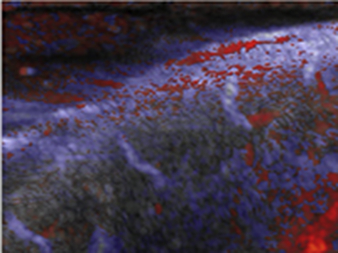
Figure 2.
Ultrafast Color Doppler Imaging of Intramural Coronary Vasculature in Open-Chest Swine Experiments
Red indicates blood flows moving upward, and blue indicates blood flows moving downward. (Top) An example of long-axis results in 1 animal: (A) venous coronary flow in mid-systole and (C) arterial coronary flow in mid-diastole. (Bottom) Results in a mid-level short-axis view showing (B) venous and (D) arterial flow in the same animal. Inversion of venous and arterial flow patterns is clearly visible: in systole, venous blood flow moves upward from the endocardium to the epicardium before moving downward in the epicardial veins. In diastole, arterial blood flow goes up in the epicardial vessels before flowing down in the myocardium. The epicardial and arteriolar compartments are successfully detected. Vessels below 100 μm remain below resolution. See Online Video 1. Scale bar = 3 mm.


