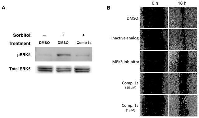
Figure 3. Activity of Compound 1s in cell-based assays.
(A) MDA-MB-231 cells were treated with DMSO or Compound 1s (10 μM) and then stimulated with sorbitol, as indicated. Cell lysates were analyzed by western blot for phosphoERK5 (pERK5) and total ERK5. Data are representative of two independent experiments. (B) A cell migration “scratch” assay was employed using MDA-MB-231 cells. After scratching the cell monolayers, cells were treated at 0 h with 1% DMSO (solvent), an inactive analog of compound 1, the MEK5 inhibitor BIX02188 or Compound 1s (10 or 1 μM). Photographs show the scratch at 0 and 18 hr as indicated. White areas are cell monolayers, while black areas are zones without cells. Each condition was performed in duplicate or singlet wells and data are representative of four independent experiments.

