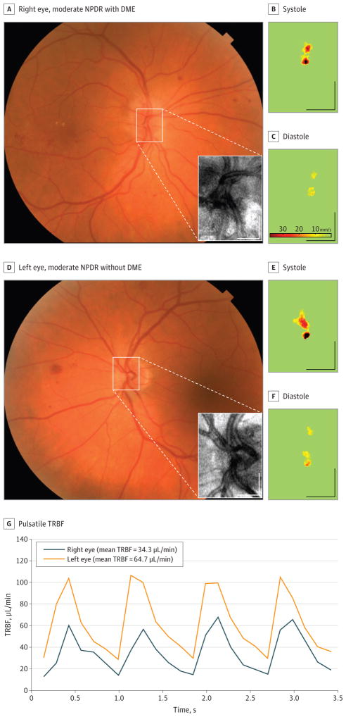
Figure 1. Measurement of Total Retinal Blood Flow (TRBF) in a Patient With Unilateral Diabetic Macular Edema (DME).
A and D, The area of the en face Doppler optical coherence tomographic scan (inset) is marked on the color fundus photograph. B, C, E, and F, Axial flow velocity en face profiles at systole and diastole. G, Pulsatile TRBF in the 2 eyes. Scale bars indicate 500 μm.

