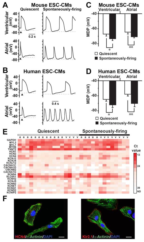
Figure 3. Spontaneously-firing m/hESC-CMs had a more hyperpolarized maximum diastolic potential (MDP).
A–B) Representative APs of quiescent and spontaneously-firing mouse (A) and human (B) ESC-CMs. C–D) The MDP of spontaneously-firing cells were significantly more hyperpolarized than that of quiescent cells in mouse (C) and human (D) ESC-CMs. *p<0.05, **p<0.01. E) A heat map of single cell qRT-PCR shows gene expression in individual atrial (a) and ventricular (v) hESC-CMs of our patch-clamp recording. F) Immunocytochemistry showed that hESC-CMs formed typical sarcomere structure and expressed HCN4 and Kir2.1. Bars indicate 10 μm.

