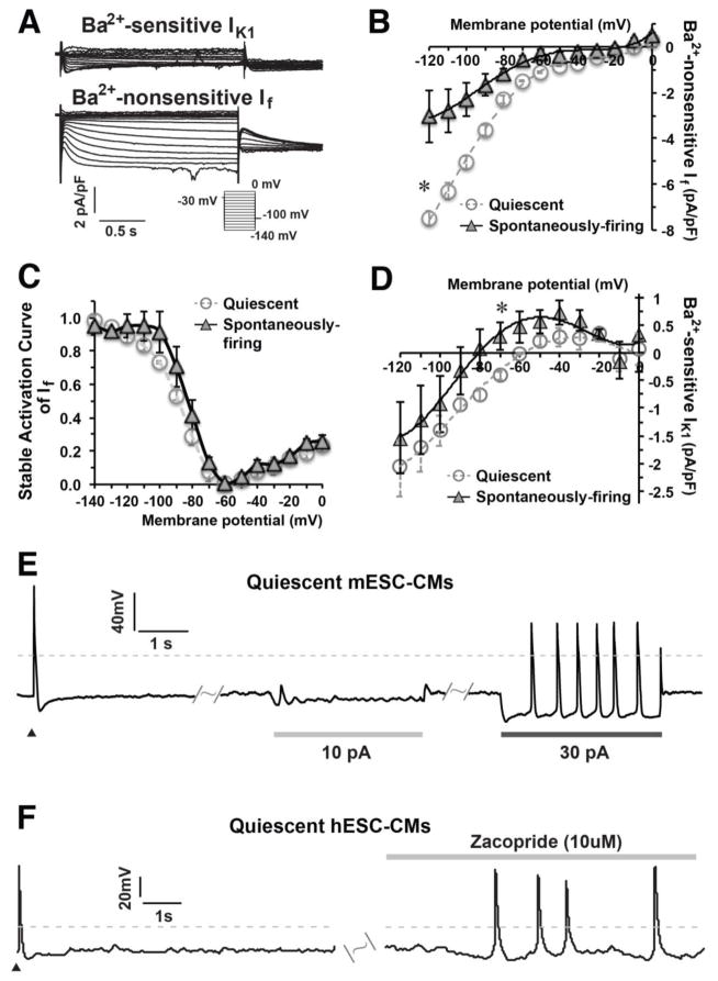
Figure 4. A hyperpolarized MP enhanced the automaticity of quiescent m/hESC-CMs.
A) Representative IK1 and If were recorded in ventricular mESC-CMs. B–C) The I–V curve and stable activation curve of If in quiescent (n=5) and spontaneously-firing (n=4) ventricular mESC-CMs. D) The I–V curve of IK1 showing that spontaneously-firing ventricular mESC-CMs (n=4) displayed a bigger outward current of IK1 than quiescent-yet-excitable ventricular mESC-MCs (n=5). E) Quiescent mESC-CMs (n=5) could spontaneously elicit APs when a proper amount of electrode-injected outward current hyperpolarized the MP. F) The automaticity of quiescent hESC-CMs (6 out of 8, right) was enhanced to fire APs spontaneously after zacopride treatment. Dash lines indicate 0 mV, ▲ indicates an electrical stimulation.

