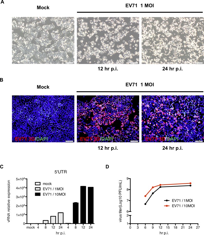
Fig 1. HT-29 cells are permissive to EV71 infection.
(A) HT29 cells were seeded on culture plates and infected with EV71 at the MOI of 1. Cell morphology was observed using an inverted microscope (magnification = 200x). (B) To confirm infection, the cells were fixed and reacted with a primary anti-EV71 3D antibody. A PE-conjugated anti-mouse IgG antibody was then applied. DAPI was used to stain the cell nuclei (magnification = 200x). (C) Total RNA was extracted from mock-infected and EV71-infected cells, and RT-qPCR was performed to detect the quantity of viral RNA. (D) Total cell lysates were harvested to detect the viral titers using a plaque assay.

