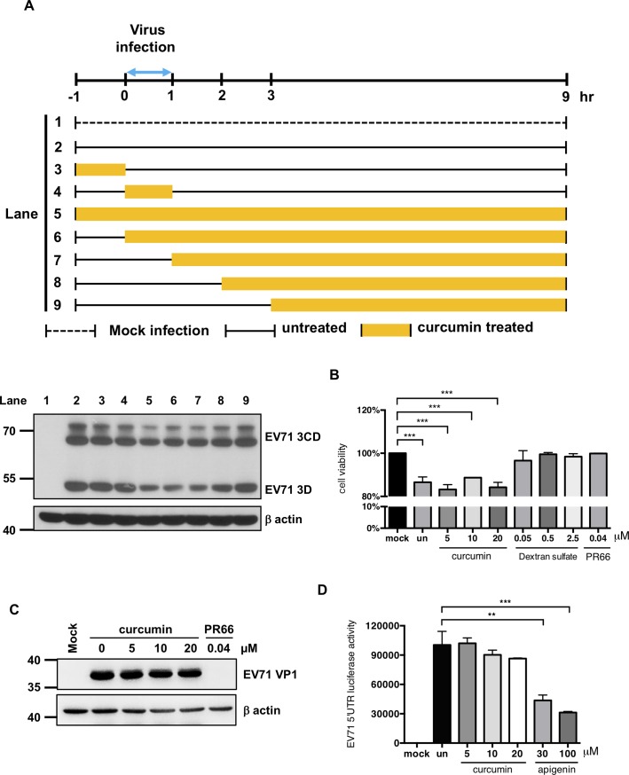
Fig 4. Curcumin inhibits EV71 protein expression when added at the early stages of infection.
(A) A time-of-addition assay was performed to assess the anti-EV71 mechanism of curcumin. HT29 cells were seeded on culture plates and infected with EV71 at the MOI of 1. Viral adsorption was allowed to occur from 0 to 1 hour p.i. Curcumin (10 μM) was added to the cells at the indicated times. After treatment, the cells were washed three times, and the medium was replaced. After 9 hours of infection, the cells were harvested, and total protein was isolated for Western blot analysis. An anti-EV71 3D antibody was used to detect the viral protein expression level. Expression of β-actin was used as an internal control. (B) To test whether curcumin can modulate the virus adsorption to the cells, EV71 virus infected cells in the absence or presence of curcumin, dextran sulfate or PR66 for one hour on ice. And cell survival rates were determined at 48 hours p.i. by MTT assay. (C) To test whether curcumin can modulate the EV71 viral particles binding to cells, EV71 were incubated with different concentrations of curcumin, or PR66 at 0.04 μM for 1 hour on ice and then used to infect the HT29 cells. Total protein was extracted at 12 hours p.i. and subsequently subjected for Western blot to examine the expression levels of viral protein VP1 and host β-actin. (D) To test whether curcumin could modulate the activity of EV71 5’UTR IRES, in vitro translation assay was used to detect the EV71 5’UTR IRES luciferase activity in the absence or presence of curcumin or apigenin. Un: untreated.

