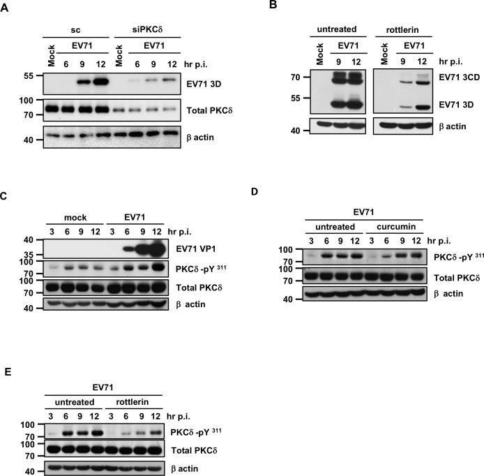
Fig 6. Curcumin inhibits EV71 infection-induced PKCδ phosphorylation.
(A) PKCδ siRNA and scramble siRNA were transfected into HT29 cells for 24 hours, and the cells were then infected with EV71 at the MOI of 1. Total protein samples were isolated at different time points and subjected to Western blot analysis to examine the expression levels of PKCδ and EV71 viral protein 3D. (B) HT29 cells were seeded and infected with EV71 in the presence of rottlerin (5 μM), a known PKCδ inhibitor. The expression levels of viral protein 3D and β-actin were then analyzed by Western blot. (C) Protein samples were harvested from mock-infected and EV71-infected HT29 cells at various time points. Immunoblotting was performed to examine the expression levels of viral protein VP1, total PKCδ, P-PKCδ (Tyr311) and β-actin. (D) Total protein was extracted from EV71-infected HT29 cells in the presence or absence of 10 μM curcumin. The expression of PKCδ, P-PKCδ (Tyr311), EV71 3D, and β-actin was analyzed by Western blot. (E) Cells were treated with rottlerin and infected with EV71. Western blot analysis was performed to detect the expression levels of P-PKCδ (Tyr311), Total PKCδ and β-actin.

