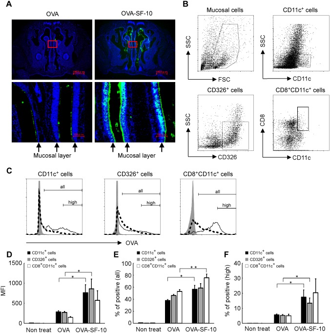
Fig 1. SF-10 enhances Ag delivery to the nasal mucosa.
Alexa647-labeled OVA or Alexa647-labeled OVA-SF-10 were instilled intranasally in BALB/c mice. Cryosections of nasal tissues were prepared 15 min after immunization (A). Top panels: representative distribution of Alexa647-labeled OVA (green) in the nasal cavity, bottom panels: high enlargement of the red square in the top panels. Nuclei stained with Hoechst33342 (blue). Arrow: nasal mucosal layer. After administration of Alexa647-labeled OVA and Alexa647-labeled OVA-SF-10 for 60 min, mucosal cells in the nasal cavity were isolated and Alexa647-labeled OVA incorporated cells in CD11c+, CD326+, and CD8+CD11c+ cells were analyzed by flow cytometry (B-F). The gating strategy of each cell population in the nasal cavity is shown (B). Alexa647-labeled OVA incorporated cells in each cell fraction from non-treated mice (gray), OVA- (dashed line) and OVA-SF-10- (solid line) immunized mice are shown (C). “all” represents all Alexa647-labeled OVA incorporated cells and “high” represents high fluorescent intensity Alexa647-labeled OVA incorporated cells. After administration for 60 min, the mean fluorescent intensity (MFI) of Alexa647-labeled OVA in each population was analyzed by flow cytometry (D) (n = 5). Each bar represents the mean ± SEM of MFI of Alexa647-labeled OVA. The percentages of positive cells in all Alexa647-labeled OVA incorporated cells and percentages of high fluorescent intensity cells that incorporated large amounts of Alexa647-labeled OVA are shown in (E) and (F), respectively, (n = 5). *P < 0.05, **P < 0.01.

