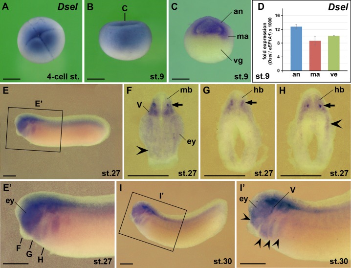
Fig 6. Dsel is expressed maternally and in the eye, brain and cranial neural crest.
Embryos after whole-mount in situ hybridization are shown in animal view (A), lateral view (B,E,E’,I,I’), hemi-sectioned (C) and transversally sectioned (F-H). (A) 4-cell stage embryo. (B,C) Blastula embryos. The bold line indicates the level of section in C. (D) qPCR analysis at stage 9. Note ubiquitous expression of Dsel in the animal cap, marginal zone and vegetal region. (E-I’) Tailbud embryos. The bold lines indicate the level of sections in F-H. Note Dsel expression in the eye, interneurons of the mid- and hindbrain (arrows), cranial neural crest (indented arrowheads) and bilateral trigeminal ganglia. an, animal cap; ey, eye; hb, hindbrain; ma, marginal zone; mb, midbrain; V, trigeminal ganglion, vg, vegetal region. Scale bars are 500 μm.

