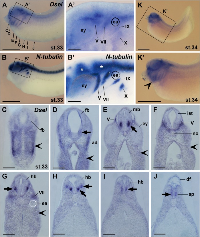
Fig 7. Dsel is expressed in differentiated neurons, cranial sensory ganglia and the spinal cord.
Embryos are shown in lateral view (A-B’,K,K’) and transversal vibratome sections (C-J). (A,A’) Embryo at stage 33. The bold lines indicate the level of sections in C-J. Magnification in A’ depicts Dsel mRNA in cranial sensory ganglia: V, trigeminal ganglion; VII, geniculate ganglion; IX, petrosal ganglion; X, nodose ganglion. (B,B’) Sibling embryo depicting N-tubulin expression in the central nervous system (stars) and cranial ganglia. (C-J) Dsel is expressed in the adenohypophysis and distinct neurons (arrows) of the forebrain, midbrain, hindbrain and spinal cord. The indented arrowhead labels signals in migrating cranial neural crest cells. (K,K’) Embryo at stage 34. The indented arrowhead labels robust Dsel transcripts in post-migratory cranial neural crest cells. ad, adenohypophysis; df, dorsal fin; ea, ear; ey, eye; fb, forebrain; hb, hindbrain; ist, isthmus; IX, petrosal ganglion; mb, midbrain; no, notochord; pm, sp, spinal cord; V, trigeminal ganglion; VII, geniculate ganglion; IX, petrosal ganglion; X, nodose ganglion. Scale bars are 1 mm (A,B,K,K’) and 200 μm (A’,B’,C-J).

