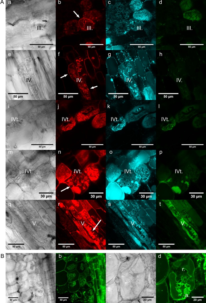
Fig 8. Localization of MtDefMd1-mGFP6 and additional fluorescence marker proteins in mycorrhized M. truncatula roots.
Differential interference contrast micrographs of razor blade hand-cut sections of transgenic M. truncatula roots are shown (A; a, e, i, m, and q; B; a and c). Confocal microscopy was used to localize a fusion of the signal peptide of MtBcp1 with mCherry under the control of the MtBcp1 promoter (A; b, f, j, n, and r), an ER-CFP fusion under the control of the 2x35S-promoter (A; c, g, k, o, and s), and an MtDefMd1-mGFP6 fusion under the control of the MtDefMd1 promoter (A; d, h, i, p, and t). Additionally, a tonoplast membrane-directed GFP fusion under the control of a 2x35S-promoter was studied (B; b and d). Roots were mycorrhized with R. irregularis for six weeks. Arrows in the MtBcp1SP-mCherry micrographs indicate structures referred to in the text. Abbreviations: a., arbuscule branches; r., restructuring; III, bird's-feet arbuscule; IV, active arbuscule; IVt, fully developed arbuscule prior to collapsing; V, collapsed arbuscule.

