
Figure 1.
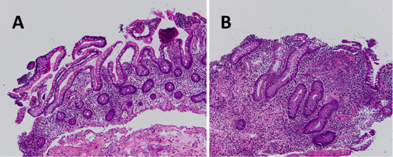
Histological Comparison of Normal Ileal Tissue and Pouchitis.
Histological representations of pouchitis are displayed. (a) 20x; Normal tissue taken during pouchoscopy at 50cm within the terminal ileum above the ileal pouch. No pathogenic alterations or inflammation is seen. (b) 20x; Biopsy obtained during pouchoscopy at 20cm within the ileal pouch showing increased acute inflammatory cell infiltration and ulceration of the epithelium, suggestive of pouchitis. The presence of blunted villi and crypt lengthening indicates significant colonic metaplasia.