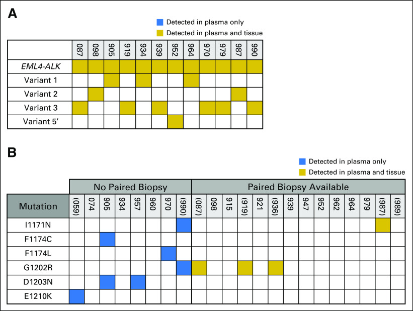
Fig 3.
Tissue–plasma concordance analysis. Gold box indicates that an alteration was detected in both plasma and tissue; blue box indicates that an alteration was present in the plasma only. (A) Concordance between fusion calls. In instances where the ALK fusion partner was not specified during tissue genotyping, patients were excluded from the concordance analysis. The fusion variant in MGH905's plasma was not identified by tissue analysis.(B) Concordance between ALK (anaplastic lymphoma kinase) mutation calls. Parentheses denote patients for whom the ALK mutation was considered the dominant resistance mechanism. For patients without paired biopsies, ALK mutations that were detected at the time of progression on an ALK tyrosine kinase inhibitor are listed. None of the patients without paired biopsies had plasma collected during progression on sequential ALK inhibitors. ALK mutations in the plasma of MGH905 occurred in cis. Because the mutations were separated by too many bases, the allelic relationship (cis v trans) could not be determined for MGH990’s ALK mutation.

