
Figure 5.
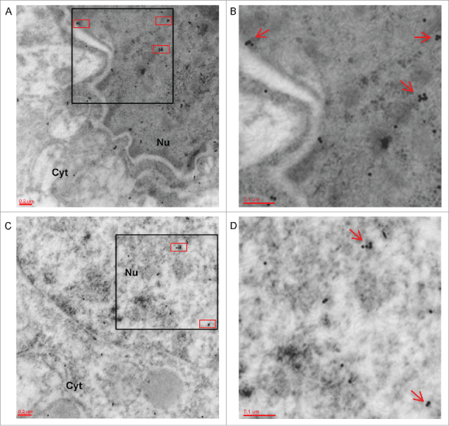
Co-localization of FMRP and ADAR2 in hippocampal sections by electron microscopy. (A-C) Two representative electronic micrographics of a double-immunogold for FMRP (labeled by 10 nm gold particles) and ADAR2 (labeled by 15 nm gold particles) performed on hippocampal ultra-thin sections of adult male mice. The red boxes highlight the co-localization of FMRP and ADAR2 proteins inside the nucleus (Nu). Magnification: 20000 x (B) 30000 x magnification of black box showed in panel A. Red arrows show FMRP/ADAR2 co-localization. (D) 30000 x magnification of black box in panel C. Red arrows show FMRP/ADAR2 co-localization. Nu: Nucleus; Cyt: Citoplasm.