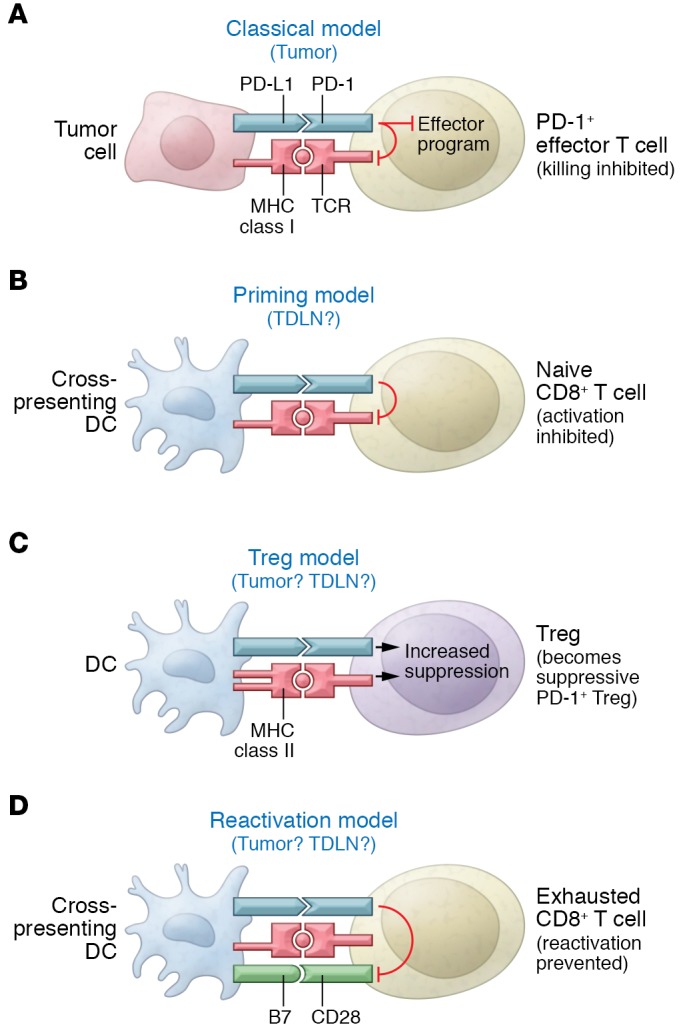
Figure 1. Possible locations for the immunosuppressive PD‑L1 targeted by checkpoint blockade.

Four different hypothetical models are presented, along with the sites in which PD‑L1 might be active, either in tumor or TDLN. (A) The traditional model, in which PD‑L1 is expressed on the tumor cell itself and directly inhibits killing of the target cell by activated PD‑1+ effector T cells. (B) Model in which inhibitory PD‑L1 is expressed on DCs during the initial priming of naive tumor-specific T cells. (C) Indirect model in which PD‑L1 delivers an activating signal to Tregs via PD‑1 and the activated Tregs then mediate immune suppression. (D) Model in which DCs in tumor or TDLNs constantly interact with mature, exhausted effector T cells and PD‑L1 serves to inhibit reactivation driven by B7-CD28 costimulation. In the figure, PD‑L1 is depicted as being expressed on the same DC that could reactivate the exhausted T cell, but this interaction might also occur in trans (PD‑L1 might be expressed on a neighboring macrophage or other APC).
