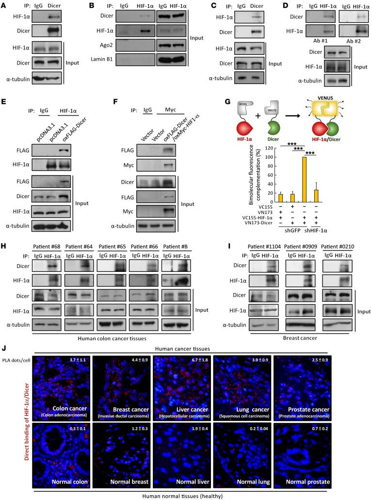
Figure 1. HIF-1α interacts with Dicer in multiple human cancer cell lines and tumors.
(A–D) Endogenous interaction between HIF-1α and Dicer. Immunoprecipitation of endogenous HIF-1α and Dicer were performed using anti–HIF-1α and anti-Dicer antibodies in HEK293T (A and B) and HCT116 cells (C and D). Ago2 was detected in anti–HIF-1α immunoprecipitates from HEK-293T cells. Lamin B1 was used as a loading control (B). (E and F) Immunoprecipitation analysis of the association between endogenous HIF-1α (E) or Myc–HIF-1α (F) and FLAG-Dicer in HCT116 cells. (G) BiFC of the association of VC115-fused HIF-1α and VN173-fused Dicer (top). HCT116 cells were transfected with VC155–HIF-1α, VN173-fused Dicer, or corresponding vector. (H–J) HIF-1α interacts with Dicer in tumor tissues. Anti–HIF-1α immunoprecipitates were isolated from lysates of surgically resected tumors to detect the in vivo interaction between HIF-1α and Dicer in colon (H) and breast (I) cancer tissues. The immunoblots presented were derived from replicate samples run on parallel gels (I). HIF-1α and Dicer were detected using in situ PLA in human colon, breast, lung, liver, and prostate normal and cancer tissues (J). PLA signals are shown in red along with DAPI nuclear staining (blue). Each red fluorescent dot indicates the direct binding of the HIF-1α/Dicer complex in close distance (<40 nm). Tissues stained with only anti–HIF-1α antibodies were also analyzed as negative controls shown in Supplemental Figure 1I. Data are presented as mean ± SD, with at least n = 3 per group. Multigroup comparisons were analyzed by 1-way ANOVA with Tukey’s post hoc test. ***P < 0.001.

