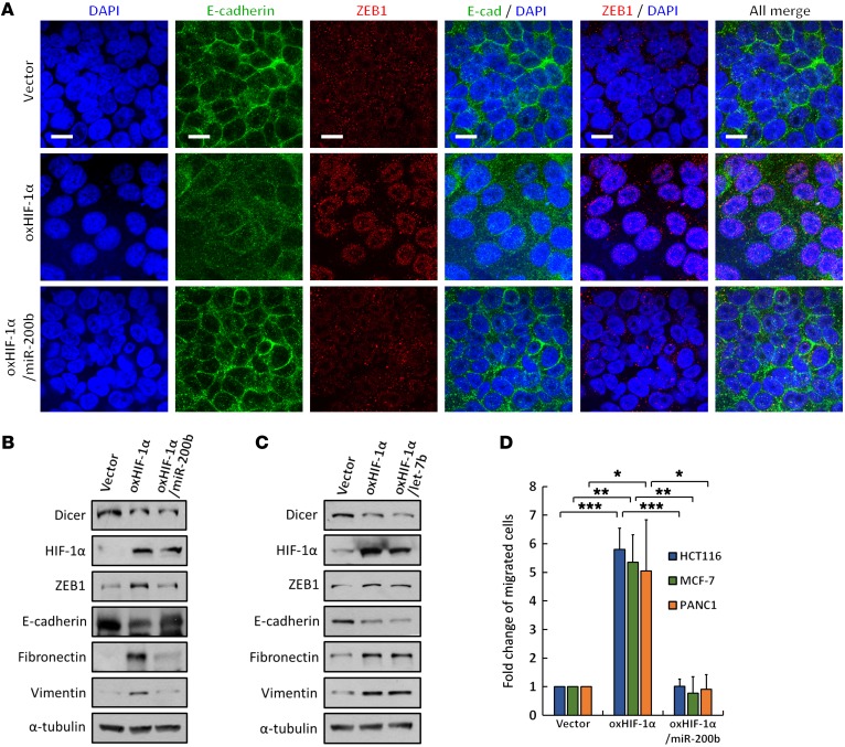
Figure 8. Functional roles of HIF-1α–induced miR-200b suppression in EMT.
(A–D) Roles of HIF-1α in miR-200b and let-7b–mediated EMT and cell migration. HIF-1α and miR-200b or let-7b were coexpressed in HCT116 cells. ZEB1 and E-cadherin were analyzed through immunofluorescence staining (A) using anti–E-cadherin and anti-ZEB1 antibodies or Western blotting (B and C) and using anti–E-cadherin, anti-ZEB1, anti-fibronectin, and anti-vimentin antibodies. Immunofluorescence images were obtained by using confocal microscopy, as indicated: E-cadherin (green); ZEB1 (red); DAPI (blue). Scale bars: 10 μm. The effects on cell migration in HCT116, MCF-7, and PANC1 cells were measured in Boyden chamber assays (D). The immunoblots presented were derived from replicate samples run on parallel gels (B and C). Data are presented as mean ± SD, with at least n = 3 per group. Multigroup comparisons were analyzed by 1-way ANOVA with Tukey’s post hoc test. *P < 0.05; **P < 0.01; ***P < 0.001.

