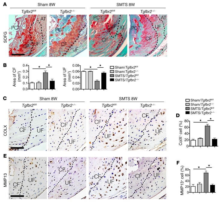
Figure 10. Deletion of Tgfbr2 in Nestin+ cells mitigates enthesis degradation in enthesopathy mice.
(A) SOFG staining of Achilles tendon enthesis. PCT, CF, UF, and Achilles tendon are separated by dotted lines. Scale bar: 200 μm. (B) Quantitative analysis of area of CF and UF. (C and E) Immunohistochemical and (D and F) quantitative analysis of (C and D) COLX+ and (E and F) MMP13+ cells in enthesis fibrocartilage. Scale bars: 150 μm. Data shown as mean ± SEM. n = 10. *P < 0.05 compared between or to the sham group.

